
Protein Labeling Reagents


Biotium CD1a(CB-T6), CF488A conjugate, 0.1mg/mL
At least five CD1 genes (CD1a, b, c, d, and e) are identified. CD1 proteins have been demonstrated to restrict T cell response to non-peptide lipid and glycolipid antigens and play a role in non-classical antigen presentation. CD1a is a non-polymorphic MHC Class 1 related cell surface glycoprotein, expressed in association with Beta-2 microglobulin. Anti-CD1a labels Langerhans cell histiocytosis (Histiocytosis X), extranodal histiocytic sarcoma, a subset of T-lymphoblastic lymphoma/leukemia, and interdigitating dendritic cell sarcoma of the lymph node. When combined with antibodies against TTF-1 and CD5, anti-CD1a is useful in distinguishing between pulmonary and thymic neoplasms since CD1a is consistently expressed in thymic lymphocytes in both typical and atypical thymomas, but only focally in 1/6 of thymic carcinomas and not in lymphocytes in pulmonary neoplasms. Anti-CD1a is reported to be a new marker for perivascular epithelial cell tumor (PEComa).Primary antibodies

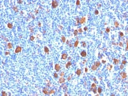
Biotium Fascin-1(FSCN1/416), CF488A conjugate, 0.1mg/mL
Recognizes a protein of 55 kDa, which is identified as fascin-1. Its actin binding ability is regulated by phosphorylation. Antibody to fascin-1 is a very sensitive marker for Reed-Sternberg cells and variants in nodular sclerosis, mixed cellularity, and lymphocyte depletion Hodgkin's disease. It is uniformly negative in lymphoid cells, plasma cells, and myeloid cells. Fascin-1 is also expressed in dendritic cells. This marker may be helpful to distinguish between Hodgkin lymphoma and non-Hodgkin lymphoma in difficult cases. Also, the lack of expression of fascin-1 in the neoplastic follicles in follicular lymphoma may be helpful in distinguishing these lymphomas from reactive follicular hyperplasia in which the number of follicular dendritic cells is normal or increased. Antibody to fascin-1 has been suggested as a prognostic marker in neuroendocrine neoplasms of the lung as well as in ovarian cancer. Fascin-1 expression may be induced by Epstein-Barr virus (EBV) infection o
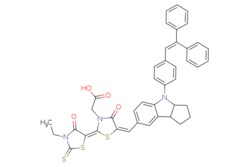
eMolecules 786643-20-7 | Medchem Express | D149 Dye | 50mg | 446271272 | HY-50938 | MFCD28016704 | 741.94 | C42H35N3O4S3
Medchem Express | D149 Dye | 50mg | 446271272 | HY-50938 | 786643-20-7 | MFCD28016704 | 741.940 | C42H35N3O4S3

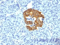
Biotium Insulin(2D11-H5), CF568 conjugate, 0.1mg/mL
Recognizes a polypeptide which is identified as insulin, a 51-amino acid polypeptide composed of A and B chains connected through the C-peptide. Proinsulin, which has very little biological activity, is cleaved by proteases within its cell of origin into the insulin molecule and the C-terminal basic residue. Insulin enhances membrane transport of glucose, amino acids, and certain ions. It also promotes glycogen storage, formation of triglycerides, and synthesis of proteins and nucleic acids. Deficiency of insulin results in diabetes mellitus. The main storage site for insulin is the pancreatic islets. Antibodies to insulin are important as beta-cell and insulinoma marker.Primary antibodies are available purified, or with a selection of fluorescent CF Dyes and other labels. CF Dyes offer exceptional brightness and photostability. Note: Conjugates of blue fluorescent dyes like CF405S and CF405M are not recommended for detecting low abundance targets, because blue dyes have



LICOR IRDye 800CW NHS Ester (1g)
IRDye infrared dyes from LI-COR Biosciences are available for researchers developing applications using near-infrared (NIR) fluorescence. These unique dyes are used in a variety of NIR imaging applications including Western blotting, plate-based assays, protein arrays, tissue section imaging, and molecular activity measurements. Standard NHS ester chemistry is used to produce custom probes labeled with LI-COR IRDye infrared dyes. The NHS ester reactive group provides the functionality for labeling primary and secondary amines, such as lysine residues in proteins.
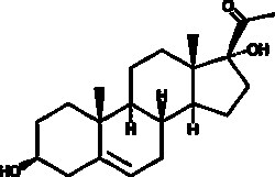
Cayman Chemical 17a-hydroxy Pregnolon 1g
A metabolite of pregnenolone; a precursor in the biosynthesis of DHEA; plasma levels are elevated in patients with type II 3β-hydroxysteroid dehydrogenase deficiency

Lumiprobe Sulfo-Cyanine5 azide, 5 mg
Water-soluble Cyanine5 dye azide for Click chemistry. Bright and photostable dye with red fluorescence. The reagent has high water solubility. Due to high solubility and hydrophilicity, the dye can be used for labeling biomolecules in native, purely aqueous conditions.
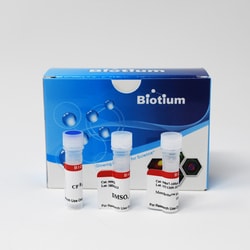
Biotium MemBrite™ Fix 640/660 Cell Surface Staining Kit, 100 Reactions
MemBrite™ Fix Cell Surface Staining Kits are designed for covalently staining the surface of live cells to conveniently visualize cell boundaries in immunofluorescence experiments. MemBrite™ Fix are new covalent cell surface stains that react irreversibly with cell surface proteins for staining that can withstand formaldehyde or alcohol fixation, and detergent permeabilization. Unlike lectins such as WGA, which bind specific targets that may vary between cell types, MemBrite™ Fix dyes react widely with cell surface proteins. MemBrite™ Fix staining is rapid and non-toxic to cells, and because MemBrite™ Fix dyes are highly water soluble, they stain cells much more evenly than traditional lipophilic membrane dyes like DiO, DiI, PKH, Vybrant, or CellMask™. This trial size kit has reagents sufficient for 100 labeling reactions. The kit contains 1 vial of MemBrite™ Fix 640/660 that is sufficient for 20 uL of 1000X dye after DMSO reconstitution.
Biotium CD4(EDU-2), CF488A conjugate, 0.1mg/mL
Recognizes a protein of 55 kDa, identified as CD4. It is a membrane glycoprotein of T lymphocytes that interacts with major histocompatibility complex class II antigens and is also a receptor for the human immunodeficiency virus. This protein is expressed not only in T lymphocytes, but also in B cells, macrophages, and granulocytes. It is also expressed in specific regions of the brain. The protein functions to initiate or augment the early phase of T-cell activation, and may function as an important mediator of indirect neuronal damage in infectious and immune-mediated diseases of the central nervous system. Multiple alternatively spliced transcript variants encoding different isoforms have been identified. This MAb was characterized as human CD4 antibody at II and IV International Workshop on Human Leukocyte Differentiation Antigens.Primary antibodies are available purified, or with a selection of fluorescent CF Dyes and other labels. CF Dyes offer exceptional brightn
Biotium CD63(LAMP3/968), CF568 conjugate, 0.1mg/mL
This MAb recognizes protein of 26 kDa-60 kDa, which is identified as CD63. Its epitope is different from that of MAb LAMP3/529. The tetraspanins are integral membrane proteins expressed on cell surface and granular membranes of hematopoietic cells and are components of multi-molecular complexes with specific integrins. The tetraspanin CD63 is a lysosomal membrane glycoprotein that translocates to the plasma membrane after platelet activation. CD63 is expressed on activated platelets, monocytes and macrophages, and is weakly expressed on granulocytes, T cell and B cells. It is located on the basophilic granule membranes and on the plasma membranes of lymphocytes and granulocytes. CD63 is a member of the TM4 superfamily of leukocyte glycoproteins that includes CD9, CD37 and CD53, which contain four transmembrane regions. CD63 may play a role in phagocytic and intracellular lysosome-phagosome fusion events. CD63 deficiency is associated with Hermansky-Pudlak syndrome and is strongly
Biotium CD11b(M1/70), CF568 conjugate, 0.1mg/mL
CD11b is a cell adhesion molecule that acts as a receptor for cell surface ligands such as intracellular adhesion molecules (ICAMs) or soluble ligands. Integrins are heterodimeric proteins that contain an alpha chain and beta chain. IntegrinαM combines with the Integrinβ2 to form a leukocyte-specific integrin referred to as macrophage receptor 1 (Mac-1), or inactivated-C3b (iC3b) receptor 3 (CR3). IntegrinαM/β2 is important in the adherence of neutrophils and monocytes to stimulated endothelium, and also in the phagocytosis of complement coated particles. The protein CD11b has been implicated in the various adhesion-related interactions of cells such as monocytes, macrophages, natural killer (NK) cells, and granulocytes. It is part of a heterodimer that consists of CD11b and CD18. It also modulates the uptake of complement-coated particles within the cell. It is commonly used as a microglial marker in tissues derived from the nervous system.Primary antib